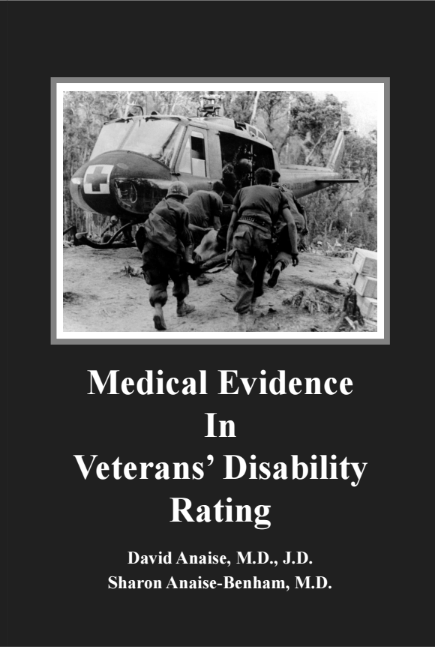
LAPAROSCOPIC CHOLECYSTECTOMY


David Anaise MD JD

Please visit my site for other articles


OPEN V. LAPAROSCOPIC CHOLECYSTECTOMY


Laparoscopic cholecystectomy first performed in 1985, has gained acceptance as the standard of care for patients requiring cholecystectomy. A NIH Consensus Development Conference held in September, 1992 concluded that "laparoscopic cholecystectomy provides a safe and effective treatment for most patients with symptomatic gallstones. Indeed, it appears to have become the procedure of choice for many of these patients." Laparoscopic surgery is popular with patients and insurance companies. The absence of an incision is cosmetically appealing. The immediate post operative course is smoother, allowing for early discharge from the hospital and early return to work. The procedure however is surgically demanding and introduces specific risks unique to the laparoscopic surgery that are not present during the performance of open cholecystectomy.

The wide-spread use of laparoscopic cholecystectomy is associated with an increased risk of intra-operative injury involving the bile ducts, bowel, and vascular structures as compared to open cholecystectomy. ( see also discussion of these injuries in my early article ) Since surgeons are reluctant to publish their own complications rate, and since the complications of laparoscopic cholecystectomy are treated in tertiary centers, the precise magnitude of the problem remains obscure.  Large population-based studies have ,however, allowed an accurate estimate the magnitude of the increased risks following laparoscopic cholecystectomy.

Reliable population based studies were conducted in Ontario Connecticut and most recently in Western Australia. The latter was recently addressed in an editorial published in the prestigious publication, Annals of Surgery. Western Australia presents ideal advantage for population-based outcome studies of surgical procedures. The population is geographically isolated, with low emigration rate and thus loss to follow up is minimal. Furthermore its capital city, Perth, is the only city large enough to support tertiary referral centers. And thus all complications caused by laparoscopic cholecystectomy and open cholecystectomy were reported.  The Australian authors after adjusting for age, gender, teaching hospital, and coexisting severe disease, found that laparoscopic cholecystectomy was 2.5 times more likely to result in intra-operative injury than an open cholecystectomy. Earlier reports expressed hope that as most surgeons passed through the learning curve of laparoscopic cholecystectomy, the incidence of injury would decline. Unfortunately a recent report of more than 10,000 cases at the military institutions have shown no significant improvement over the initial report from the same institutions.

Misidentification of anatomy appears to be the most common cause of laparoscopic bile duct injury. The most common scenario, initially described by surgeons at Duke University as the "classic" Injury, occurred in 63% of their patients. The "classic" injury occurs due to mistaking the common bile duct for the cystic duct. The Common Bile Duct then is clipped and divided. Further retraction of the gallbladder then leads to a second higher injury with division of the common hepatic duct as it approaches the bifurcation. This second ductal injury is often described in the operative note as being a 'second cystic duct" or "an accessory duct."  This injury is particularly devastating as complete transection of the biliary tree virtually removes any possibility of non-operative management by either endoscopic or transhepatic technique and mandates the need for surgical reconstruction of the biliary tree to establish biliary-enteric drainage.

Other less common mechanisms of injury include a "tenting injury" in which the common bile duct is pulled laterally at the time of occlusion of the cystic duct and caught in the clip, thermal injuries due to injudicious use of cautery or laser, excessive application of clips to control bleeding in the area of the triangle of Calot. and. finally, injuries to an aberrant or low-inserting right hepatic duct. Regardless of the nature of the injury, the majority of. biliary injuries are not recognized during the initial laparoscopic cholecystectomy. In their initial series, the Duke surgeons found that in all 12 patients with major bile duct injuries, the injury was not recognized at the time of laparoscopic cholecystectomy. Lillemoe of John Hopkins reported that fewer than one third of injuries were recognized at the original operation. Delay in recognition of biliary injuries invariably results in infection and significantly reduces the chance for optimal outcome after reconstruction.

STANDARD OF CARE


The mere fact that the common duct was injured during the performance of laparoscopic cholecystectomy is not by itself sufficient to establish liability. Nor does the fact that the surgeon chose to perform laparoscopic cholecystectomy rather than open cholecystectomy create a presumption of deviation from the standard of care. Rather, there is a presumption that the failure to adhere to established steps necessary to prevent such injuries breaches the surgeons duty to his patient.

The Society of American Gastrointestinal Endoscopic Surgeons is the leading professional society representing more than 3000 board certified surgeons who use endoscopy and laparoscopy as an integral part of their treatment of patients. The society established guidelines for the clinical application of laparoscopic biliary tract surgery. The Guidelines are based in turn on an important 1991 publication by Hunter who described the steps a surgeon must take in order to avoid this dreadful complication. The guidelines require the surgeons to adhere the the following steps:

  • The surgeon must clearly identify the cystic duct at its junction with the gallbladder
  • The surgeon should retract the gallbladder infundibulum laterally rather than in cephalad direction
  • The surgeon should Meticulously dissect the cystic duct and cystic artery.
  • The surgeon should limit the use of all energy sources near the Common Bile Duct and recognize that they can cause occult injury.
  • The surgeon should use operative cholangiography liberally to discover surgically important anomalies, clarify difficult anatomy and to detect common bile duct stones.
  • The surgeon should not hesitate to convert to an open operation for technical difficulties, anatomic uncertainties, or anatomic anomalies, especially in cases of acute cholecystitis
  • The surgeon need to see all structures clearly before dividing any ductal structures

Lateral retraction of the infundibulum

In 1991 Hunter proposed a 5 step approach to prevent the high rate of biliary injury in the United States. Hunter noted that Bile duct injuries with laparoscopic cholecystectomy appeared to be more common in the U.S. (0.5 to 2.7%) than in Europe (0.33%) . He observed that American teaching stressed cephalic (towards the Right shoulder) traction on the infundibulum of the gallbladder, tenting the CBD and risking its mis-identification. Figure 1 shows the normal relationship between the cystic duct and the Common Bile Duct . The cystic duct emerges at acute angle to the CBD and this angle actually narrows when the fundus is retracted towards the shoulder. From the perspective of the telescope, the distal Common Bile Duct appears continuous with the cystic duct and can easily be mistakenly identified as a long cystic duct. European instruction stresses the lateral retraction depicted in Figure 2 Such retraction places the cystic duct at right angles to the CBD, reducing the likelihood of misidentification. Figure 3 depicts the preferred retraction of the fundus and infudibulum as videotaped in actual laparoscopic operation and correlate with steps 1-3 in Hunter's original publication. It also shows the cannulation of the cystic duct in preparation for Operative Cholangiography.


Fig 1.  the cystic duct emerges in acute angle from the CBD


Fig 2.  The cystic duct is brought to a right angle with the CBD by lateral retraction of the infundibulum Hunter et al. Am J Surg 162;73 1991


Fig 3 operative picture depicts the correct retraction of the infundibulum stretching the cystic duct. Note the cystic artery parallel to the cystic duct. The Operative Cholangiography cannula

Was inserted


Meticulous dissection

Step 4 depicted in figure 4 is arguably the most important step in preventing CBD injury. No clip should be placed on, and no incision should be made in, any structure until the transition between cystic duct and gallbladder infundibulum is clearly visualized. It is not adequate to see the cystic duct '"entering" the gallbladder" as this may belie a tented Common Bile Duct coursing behind the gallbladder, drawn up by chronic inflammation. Safe dissection absolutely requires that the cystic duct must be seen widening into the gallbladder before one can certify accurate anatomic identification. In the case of the absent, or short, wide-mouthed cystic duct identified during this step or in step 5, the surgeon must consider conversion to open cholecystectomy or develop the skills to laparoscopically suture the cystic duct stump without impinging on the lumen of the Common Bile Duct .


 Fig 4 dissection of the funnel Hunter et al. Am J Surg 162;73 1991


Operative Cholangiography


Hunter's Step 5, is Operative Cholangiography. Operative Cholangiography is necessary for the detection of common bile duct stones. In 15% of the cases a surgeon finds a stone which escaped from the gallbladder and is lodged in the distal Common Bile Duct.  If left in the duct the stone often obstructs the duct causing cholangitis, Common Bile Duct stricture and pancreatitis. Operative Cholangiography is of particular importance in laparoscopic cholecystectomy because it greatly clarifies the anatomic relationship of the biliary tree. Hunter's principles emphasize the need to see everything well before cutting anything while keeping dissection away from the area where almost all biliary anomalies would be encountered: the triangle of Calot. After following these principles, an Operative Cholangiography will help confirm the anatomy before transecting any structure. Figure 5 depicts an actual , Operative Cholangiography The surgeon in this case can be satisfied that no stones are lodged in the Common Bile Duct , no injury to the Common Bile Duct occurred and that the clip on the cystic duct is not encroaching on a common "tented " Common Bile Duct.

Fig 5  Operative Cholangiogram. From  "The operation " The learning Channel
 

Of course the Operative Cholangiography must be interpreted correctly by the surgeon. Like proficiency in the technique of laparoscopic cholecystectomy itself, surgeons who routinely perform Operative Cholangiography become better in performing and interpreting the study. The Australian study reported that the risk of bile duct injury was decreased by half if Operative Cholangiography was performed during either the laparoscopic or open cholecystectomy. As case complexity increased ( e.g., severe inflammation around the gall bladder), obtaining an Operative Cholangiography decreased the risk of bile duct injury by eight-fold!.

Common Bile Duct injuries could occur despite Operative Cholangiography. A surgeon who misidentifies the cystic duct will partially cut the Common Bile Duct in order to insert the Operative Cholangiography cannula only to find out his mistake after Operative Cholangiography was performed. Partial cut of the Common Bile Duct is a minor complicationcommonly. Surgeon routinely enter the duct   in approximately 10-15% of open cholecystectomy in order to retreive  Common Bile Duct stones. It is repaired easily with no appreciable post operative morbidity. The Armed forces reported that such minimal injury occurred in 18 of the 25 patients ( 72%) who suffered Common Bile Duct injuries despite the performance of Operative Cholangiography. Another 6 patients suffered from  transection of the Common Bile Duct without clip damage and were  managed by simple direct biliary anastmosis. Only one patient of the 25 reported (4%) had complex biliary injury requiring complex repair. In contrast six of the 12 patients ( 50%) who suffered from Common Bile Duct injuries and did not have Operative Cholangiography, suffered from complex excision of the Common Bile Duct.


Repair

Defendants often claim that while injury to the duct has occurred it was immediately recognized and repaired. While the early recognition is important to prevent infection and excessive scarring, immediate repair of the Common Bile Duct is associated with unacceptably high failure and it often compounds the initial injury.

The armed forces report, discussed earlier, has classified Common Bile Duct injuries to three classes. Class one refers to simple cut of only part of the Common Bile Duct circumference. Such injuries are easy to repair with high degree of success. Class 2 injuries involve complete transection of the duct but without clipping the duct. This injury requires more complex repair and intermediate long term result. The most difficult injury and unfortunately the most common is the removal of part of the Common Bile Duct. Study from Duke University reported that the most common scenario involved mistaking the common bile duct for the cystic duct. Once the common duct was divided and retracted with the gallbladder a second higher injury resulted with division of the common hepatic duct, as it approached the bifurcation this second ductal injury was often described as an accessory duct. Sometimes the right hepatic artery was also injured in the process leading to liver infarct.

 Fig 7

Fig 7 depicts the cystic duct as it is transected between clips. If the surgeon has mistaken the Common Bile Duct to be the cystic duct an identical picture will result. The area damaged by the clips cannot be repaired and the segment damaged must be excised. The resulting gap can be bridged only with excessive tension resulting in early failure. Lillemoe reported that end-to-end ductal repair over a T-tube was unsuccessful in all of those patients in which a complete transection of the bile duct had taken place. The standard operation for reconstruction of a major bile duct injury after laparoscopic cholecystectomy is a Roux-en-Y hepaticojejunostomy.( see Fig. 8) This requires great degree of surgical skills and should be attempted only by surgeons with great experience in performing this procedure. Lillemoe reported that when the initial surgeon performed Roux en-Y hepaticojejunostomy to repair the Common Bile Duct, 63% were successful. Only few surgeons were skilled enough, however, to perform this demanding procedure. As a result attempts at repair performed by the primary surgeon were successful in only 7% of cases and in no case was a secondary repair by the original surgeon successful. In contrast in those patients in which the first repair was performed by a tertiary care biliary surgeon, a 94% success rate was obtained.

A multi-center report by Woods from the Virginia Mason Clinic in Seattle. Washington. the Lahey Clinic in Burlington. Massachusetts, and the Mayo Clinic in Rochester, Minnesota, reported a similar high rate of failure (94%) for biliary-enteric bypass performed outside of the referral center. Conversely, all patients managed by reoperation (N = 29) at the tertiary center had a successful result.
The challenges are greatest in patients with class II, class III, and class IV injuries; in patients with injuries that occurred more than a few weeks previously; and in patients with previous unsuccessful injury repairs. Scar formation in these last two situations makes the planes either difficult to identify or nonexistent. Large blood vessels are within a few millimeters of the duct, and getting adequate exposure high in the hilum of the liver may be difficult. Yet, the scar tissue at the site of injury must be mobilized, the end of the duct must be debrided of this scar tissue back to healthy tissue, and the ductal blood supply must be preserved. It is not surprising, therefore, that the ability to accomplish these technical objectives improves with experience and that the outcome of operations for bile duct injuries is so strongly related to the surgeon's previous experience. Because bile duct injuries are uncommon, the opportunity to obtain the necessary experience is largely confined to specific surgeons in tertiary care medical centers. Most experts, therefor, advise surgeons not to attempt to reconstruct complex ( class III) Common Bile Duct injuries but rather leave the clips to avoid bile spillage ,
insert a drain and transfer the patient promptly to a tertiary care facility.

Fig 8. Roux en Y choledocho-Jejunostomy 

Manual of vascular access and transplantation Springer Vernalk

DAMAGES

Benign stricture of the common bile duct is a serious complication of upper abdominal surgery, turning, if untreated, into repeated cholangitis, biliary cirrhosis, hepatic failure, and death. Duodenal ulcer has been reported to develop in some instances as a consequence of Roux-en-Y biliary enteric repair, thus representing an additional cause of late morbidity.

Due to the difficult and delicate reconstruction of Common Bile Duct injuries, it is nor surprising that the long term results are far from being satisfactory. Perhaps the longest follow-up available after the management of major bile duct transections in the laparoscopic cholecystectomy era is reported by Bergman et al from the Netherlands in which patients sustaining major bile duct injury were reconstructed with a Roux-en-Y hepaticojejunostomy. At a median follow-up of 25 months (range, 6-38 months) 33% of the patients required subsequent transhepatic balloon dilatation or reconstruction with a secondary hepaticojejunostomy.

Tocchie et al reported that the 30-day operative mortality rate was 2.2%. Complications occurred in 18 (21.4%) patients. Several patients had more than one complication. Excellent or good results were achieved in 70 (83%) patients, whereas the remaining 12 patients experienced fair or poor results.. Anastomotic strictures requiring further treatment occurred in 10 patients, in 6 within 5 years, and in 4 at 62, 75, 85, and 96 months

Bauer reported the Philadelphia experience. Over a mean follow-up period of 11.5 +/- 10.5 months, 11 patients (38%) required 19 emergency readmissions, most commonly for cholangitis. Five patients (17%) required postoperative balloon dilatation for biliary stricture. At follow-up, 18 patients (62.0%) remain asymptomatic with normal liver function, eight (28%) are experiencing episodic cholangitis, and three (10%) are asymptomatic with persistently elevated liver function values. Bauer concluded that the consequences of a major biliary tract injury following laparoscopic cholecystectomy include a complex operative repair resulting in a lengthy postoperative stay with an increased risk of death, an excessive number of perioperative diagnostic and therapeutic studies, frequent readmissions (often as emergencies), and a lifelong risk of restricture. "The "cost" to these patients remains enormous".

Laparoscopic cholecystectomy did not become wide-spread until 1992. Studies of the results of biliary reconstruction after laparoscopic cholecystectomy are therefor incomplete because they reflect only a short post -operative follow up. The longest study is limited to only 3 years. Scarring and therefor biliary stricture progress relentlessly. Pitt et al. in their analysis of factors influencing the outcome after repair of postoperative bile duct strictures in the pre-laparoscopic era have reported that only 68% of recurrent strictures will have developed by 3 years after repair. Table 1 summarizes the world experience in biliary reconstruction in the pre -laparoscopic cholecystectomy era. One of four patients outcome was considered unsatisfactory. Many required numerous surgical procedures, underwent debilitating bouts of cholangitis, developed liver cirrhosis and ultimately required liver transplantation.


Study # patients Satisfactory % Satisfactory patients
Warren 477 66 314
Bismuth 123 99 121
Pitt 138 77 106
Kalman 63 79 46
Pellegrini 55 73 40
Genest 105 93 97
Innes 22 95 20
Bottger 72 50 36
Schweizer 17 76 13
Periera 31 92 28
Raute 64 75 48
total 1167   869


Table 1 Total success 869/ 1167 = 74%


Despite improvement in surgical technology in the laparoscopy cholecystectomy era, experts believe that long term results of current biliary reconstruction will be worse than the results of biliary reconstruction in the open cholecystectomy era. As the Duke study have shown, the most common biliary complication in the laparoscopic cholecystectomy era was excision of a long segment of Common Bile Duct requiring reconstruction at the porta hepatis. Toccchi et al have shown that the outcome of reconstruction of duct injury without loss of tissue was satisfactory in 79% of the cases. In contrast reconstruction of complex biliary injury was satisfactory in only 7%-21% of the cases. Lillemoe of John Hopkins who reported excellent short term results of biliary reconstruction cautioned that " It has been suggested that laparoscopic bile duct injuries may have a less satisfactory outcome because of both the more complex nature of many of the injuries and the frequent association with significant inflammation and fibrosis secondary to the bile leakage. Furthermore, the high percentage of failed operations performed before referral to a tertiary biliary tract surgical center may lead to a poorer outcome"

Hospital liability

The hospital may be liable for negligent credentialing , proctoring and supervising the surgeon. The Society of American Gastrointestinal Endoscopic Surgeons (SAGES) recommends the following guidelines for privileging qualified surgeons in the performance of general surgical procedures utilizing laparoscopy. The basic premise is that the surgeon must have the judgement, training and the capability of immediately proceeding to a traditional open abdominal procedure when circumstances so indicate. The society Guidelines were created to to assist hospital in complying with standard JCAHO guidelines for granting hospital privileges, and to ensure that surgery is performed only by individuals with appropriate competence, thus assuring high quality patient care and proper procedure utilization.

Granting Hospital Privileges

Prerequisite training must include satisfactory completion with Board eligibility or certification from residency programs in general surgery accredited by the Accreditation Council for Graduate Medical Education or the equivalent body if the program is based outside the United States or Canada. For those surgeons without residency training which included laparoscopic surgery or without documented prior experience in these areas, the training should include didactics, hands-on experience, participation as a first assistant and performance of the operation under proctorship. The basic minimum requirements for training should be: training in laparoscopic general surgery by a surgeon experienced in laparoscopic surgery or completion of a didactic course sponsored by an institution or society accredited by ACCME.. The individual must demonstrate to the satisfaction of an experienced physician course director/ preceptor that he/she can perform a given procedure from beginning to end.. Attendance at short courses which do not provide supervised hands-on training or documentation of proficiency is not an acceptable substitute,

Proctoring

Once credentialed, proctoring by a a qualified, unbiased staff surgeon experienced in laparoscopic surgery is recommended until proficiency has been observed and documented in writing. The proctor should be responsible to the privileging committee, and not to the patient or to the individual being proctored. Documentation of the proctor's evaluation should be submitted in writing to the privileging committee. Criteria of competency for each procedure should be established in advance and should include evaluation of: familiarity with instrumentation and equipment, competence in their use, appropriateness of patient selection, clarity of dissection, safety, time taken to complete the procedure and successful completion of same.

Supervision

Hospital should be routinely involved in the ongoing renewal of privileges, there should be a mechanism for monitoring competence. This should be done through existing quality assurance mechanisms. This should include monitoring utilization, diagnostic and therapeutic benefits to patients, complications and tissue review. Continuing medical education related to laparoscopic surgery should be required as part of the periodic renewal of privileges. For the renewal of privileges an appropriate level of continuing clinical activity should be required. In addition to satisfactory performance as assessed by monitoring of procedural activity through existing quality assurance mechanisms continuing medical education relating to laparoscopic surgery should also be required.

First assistant

The first assistant is responsible for maintaining proper exposure and is indispensable member of the team.  The American College of Surgeons established the following guidelines: The first assistant to the surgeon during a surgical operation should be a trained individual who is capable of participating in the operation and actively assisting the surgeon as part of a good working team. The first assistant provides aid in exposure, hemostasis, and other technical functions, thereby helping the surgeon carry out a safe operation with optimal results for the patient. Ideally, the first assistant to the surgeon at the operating table should be a qualified surgeon or resident in a surgical education program that is approved by the appropriate residency review committee and accredited by the Accreditation Council for Graduate Medical Education. It is a principle of surgical education and care that residents at appropriate levels of training should be provided with opportunities to assist at and participate in operations. Other physicians who are experienced in assisting the responsible surgeon may participate when a trained surgeon or a resident in an accredited program is not available.

Videotaping

The operative camera is connected to a VCR which accepts regular VHS cassettes. The recorded tape records faithfully whatever the surgeon has seen and done. Surprisingly videotaping is not treated like medical record and majority of surgeons apparently through instructions by their hospitals and insurance carriers do not videotape their procedure because they believe that it will be used against them in court. A well informed jury may, however, draw the correct inference that the lack of the videotape is similar to lack of written notes in the medical chart. It was done deliberately either to hide the physician mistake or as a sloppy practice.

Surgeons who want to learn from their mistakes record their operations. Patrick Walsh, MD, director of the Brady Urological Institute published his experience in videotaping at the January 2000 issue of Urology. When complications did occur Walsh reviewed the tapes and found subtle information that allowed him to improve his technique. Walsh offered the following advise "In radical prostatectomy, minor differences in surgical techniques can have a major impact on results. Surgeons who have less than optimal results should consider videotaping every operation and, when they have a successful one, they should watch the videotapes and see what they did right."

Videotaping should be considered essential medical record. Once biliary injury occurred the precise location and magnitude of injury is of paramount importance. Rather than subjecting the patient to expensive and dangerous radiology test to identify the source of the bile leak the surgeon can get all the information needed by reviewing the tape.  Where delay in recognition of the complication occurred the scarring and infection will hinder exploration of the Common Bile Duct. Precise information as to what actually happened rather than what the perception of the surgeon was at the time is of paramount importance to plan for the proper repair

Hospitals could improve their capacity to supervise surgeons by requiring videotaping and using the tapes as a mean to proctor, supervise and credential surgeons. As this technology is available Hospital cannot defend their lack of supervision by asserting ignorance.


Social Security Disability Tucson Arizona

1


1